Fatigue Blood Panel for Perimenopausal Women
Testing multiple markers together reveals which fatigue drivers actually matter for each woman.

Fatigue in perimenopause arrives as a single complaint with a dozen possible origins. A well-constructed fatigue blood panel addresses that reality directly: it measures reproductive hormones, thyroid function, and iron stores together, so the pattern they form can reveal which drivers are actually at work. The goal is not to produce a single number but to replace guesswork with a map.
Why fatigue in perimenopause is not a single problem with a single cause
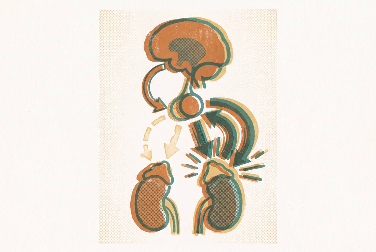
The hypothalamic-pituitary-ovarian axis, the feedback loop that governs reproductive hormones, begins shifting long before the final menstrual period. Progesterone declines first as ovulation becomes inconsistent, while estrogen becomes erratic rather than steadily lower. The body is therefore not moving through a clean descent but through a period of volatility, one in which the regulatory signals themselves are unstable. That instability, not a simple hormone deficit, is the biological substrate of perimenopausal fatigue. Cleveland Clinic describes perimenopause as a period in which the ovaries begin producing less hormones, causing cycles to become erratic or irregular. This distinction matters because an unstable system produces symptoms that fluctuate: a woman may feel profoundly exhausted one week and nearly normal the next, which makes the fatigue easy to dismiss as stress or overwork when it is in fact a direct consequence of the shifting endocrine landscape.
Sleep disruption compounds the picture. Night sweats and insomnia are documented perimenopausal symptoms driven by changing hormone levels, and they fragment restorative sleep in ways that leave a woman exhausted independent of any metabolic deficit. A night of broken sleep produces fatigue the next day even when every blood marker is perfectly normal, which means any panel that attempts to explain fatigue must account for the fact that some of that fatigue travels through the bedroom rather than through the bloodstream alone.
Mood-related fatigue adds a third layer. Estrogen influences serotonin and other mood-regulating chemicals, so its instability can produce the kind of low-grade anxiety and depressive symptoms that drain energy before a woman even gets out of bed. A 2025 study in BMC Women's Health documents a rising global burden of anxiety disorders among perimenopausal women, linked to hormonal changes affecting the body's regulatory feedback mechanisms. The fatigue, in other words, is partly metabolic, partly behavioral, and partly psychological, and these layers reinforce one another.
That raises an important question: if the drivers interact so tightly, what can a blood panel actually accomplish? The answer is that it can separate what is measurable from what is inferred. Attributing perimenopausal fatigue to "just perimenopause" or "just stress" without testing is guesswork, and testing only one marker at a time produces the same result. The panel earns its place by measuring the systems that can be measured, precisely because so much of the symptom picture resists easy attribution.
Why the overlapping conditions — thyroid dysfunction, iron deficiency, and others — must be ruled in or out at the same time
Several common conditions produce fatigue that is clinically indistinguishable from perimenopausal fatigue, and a panel that tests only reproductive hormones will miss them entirely. Hypothyroidism, including subclinical and autoimmune forms, overlaps with perimenopause on nearly every symptom: fatigue, brain fog, weight change, mood shifts, temperature sensitivity, and hair thinning. A woman experiencing all of these could reasonably conclude she is in perimenopause, and she might be right, but she might also have an underactive thyroid that is entirely treatable and entirely separate. The possibility of co-occurrence makes the problem more pressing, not less. A TSH-only order therefore produces false reassurance: the number comes back normal, and both the woman and her clinician conclude the thyroid is fine, when in fact the test simply did not look deeply enough.
Iron-deficiency anemia is the second major look-alike. A woman whose periods have become heavier may be losing iron faster than her body can replace it, and the resulting deficiency produces symptoms that layer directly on top of any hormonal fatigue. Testing ferritin, the storage form of iron, catches this problem before it is mistaken for something else.
The practical risk of attribution error is that a treatable condition goes unaddressed for months or years. The panel resolves this not by adding unnecessary tests but by including the markers that cover the most clinically relevant look-alikes in a single draw, so the picture that emerges is diagnostic rather than merely suggestive. The panel, in other words, is performing differential work, distinguishing perimenopause from its imitators rather than confirming it in isolation.
One might argue that testing for multiple conditions at once is excessive, that a focused approach is more efficient. A panel that includes thyroid and iron markers alongside reproductive hormones is not casting a wide net. It is drawing the smallest net that can actually catch the problem.
The core hormones to test and what each one reveals about fatigue
Each reproductive hormone in a perimenopause panel answers a distinct question about why the body feels the way it does, and no single marker can substitute for the pattern the full set reveals. Estradiol (E2) measures the primary estrogen circulating in the body. A single estradiol reading therefore captures a moment in a fluctuating system, which is why it must be read alongside the other markers rather than on its own.
FSH, or follicle-stimulating hormone, provides the context that makes estradiol interpretable. A trending-upward FSH alongside erratic estradiol is one of the clearest biochemical signals of the perimenopause transition, because it shows the feedback loop straining to maintain a rhythm the ovaries can no longer sustain. The two markers together reveal the dynamic, where either alone shows only a static value.
Progesterone is often the first hormonal shift in early perimenopause, because it depends on ovulation and ovulation becomes inconsistent before anything else. Low progesterone means disrupted sleep architecture, since progesterone has a calming, sleep-promoting effect via GABA pathways. When progesterone is low, estrogen also goes unopposed, an imbalance that contributes to menstrual irregularities and an increased risk of uterine lining thickening.
LH, luteinizing hormone, becomes increasingly variable as the transition progresses. DHEA-S, an adrenal androgen, contributes to energy and vitality, and declining levels can contribute to fatigue and low drive. It completes the picture by acknowledging that the adrenals, not just the ovaries, participate in the hormonal environment of midlife.
The combined picture is what matters. FSH trending upward, combined with low progesterone and erratic estradiol, alongside the symptoms themselves, constitutes a strong perimenopausal pattern that no single marker produces on its own. The panel works not because any one number is definitive but because the numbers form a pattern when read together.
The thyroid and metabolic markers that belong on the same panel
A perimenopause fatigue panel that omits thyroid and metabolic markers is structurally incomplete, because the conditions those markers detect are both common and symptomatically indistinguishable from hormonal fatigue. The thyroid sits at the center of this problem. A 2026 guide to female hormone tests notes that hormone disorders go undiagnosed or misdiagnosed among women, and that thyroid disorders are among the conditions abnormal hormone levels can indicate.
Free T4 measured alongside TSH is the pairing that makes thyroid testing meaningful. Together they distinguish primary hypothyroidism, where the thyroid itself is underperforming, from pituitary-driven dysfunction, and they catch cases where TSH alone appears normal. A woman whose TSH is unremarkable but whose Free T4 is low has a thyroid problem that a TSH-only order would have missed entirely.
Ferritin is the most sensitive marker of iron stores, and it belongs on this panel for a specific reason: heavy or irregular perimenopausal periods deplete iron faster than the body can replace it. Testing it separates the two, because a woman with both perimenopausal hormone shifts and iron deficiency has two treatable problems, not one complicated one. The treatment for each is different, and neither treatment works for the other.
SHBG, sex hormone-binding globulin, contextualizes the hormone numbers. A low SHBG can mean hormones are present but inaccessible, while a high SHBG reduces free hormone availability. Without SHBG, the estradiol value alone can mislead: a woman whose total estrogen looks adequate may still be experiencing symptoms because too little of it is free to act on tissues.
Vitamin D deficiency produces fatigue and musculoskeletal aching that compounds perimenopausal symptoms, and it is commonly low and commonly overlooked on a standard workup. This marker matters not because it is exotic but because it is so frequently abnormal and so directly tied to the complaint that brings the woman in. A panel that catches a vitamin D deficiency while also mapping the hormonal transition has done something a reproductive-only panel cannot: it has found every measurable contributor to the fatigue, not just the most obvious one.
When and how to time the blood draw so the results are actually interpretable
The same hormone panel can produce misleading results depending on where in the cycle the blood is drawn, which makes draw timing as important as marker selection. For women still cycling, days 2 through 5 of the cycle, with day 1 being the first day of the period, are the conventional recommendation for baseline reproductive hormones. A 2026 guide to female hormone tests states that testing is typically recommended on days 3 through 5, when hormone levels are at baseline, for the most accurate interpretation.
Progesterone is the exception to this rule. Its meaningful window is the mid-luteal phase, approximately days 19 through 22 of a 28-day cycle, because it peaks after ovulation. Drawing progesterone at the same time as FSH and estradiol produces an artificially low reading that may be mistaken for deficiency, when the reality is simply that the test was taken before ovulation had a chance to occur. Why exactly does this matter? Because a low progesterone reading on day 3 is normal and uninformative, while a low progesterone reading on day 21 is a genuine signal about ovulatory function.
For women with irregular cycles, which is the defining feature of perimenopause, strict day-specific timing becomes difficult. A woman who has not had a period in two months cannot wait for day 3. She draws when the cycle starts, whenever that is, and the clinician reads the result against that reality. The timing rule, in other words, must bend to the very irregularity it is meant to measure.
Thyroid, ferritin, and vitamin D are not cycle-dependent and can be drawn at any time. They do not require the same timing precision as reproductive hormones, which means a single visit can capture both the timed markers and the untimed ones without compromise. This is a practical advantage: the panel does not require multiple trips for multiple markers, only an awareness of which markers care about the calendar.
A single draw on the wrong cycle day can produce a falsely normal or falsely elevated FSH or estradiol, leading to incorrect reassurance. This is one reason why longitudinal testing across multiple draws tells a more accurate story than any single result. Timing determines whether a number is interpretable, and repetition determines whether an interpretable number is representative.
Why a single test result is rarely enough to understand perimenopausal fatigue
Hormones in perimenopause do not follow a predictable declining trajectory, which means a single snapshot test, even a well-timed one, can look normal one month and dramatically different the next. Research has demonstrated that the relationships among estrogen, LH, progesterone, and FSH evolve across transition stages. Interpreting them together and over time provides a clinical framework for understanding changing ovarian function that a single draw cannot replicate. The Swiss Perimenopause Study illustrates this directly. Serial measurement is therefore more clinically informative than single snapshots, because it captures the fluctuation itself rather than one point within it. FSH in particular is known to fluctuate from month to month during perimenopause. The trajectory of these markers over months carries information that no individual value contains. The practical value of tracking is that a woman who tests every few months accumulates a record that shows direction and velocity of change. The evidence supports repeated measurement not as a luxury but as the only approach that matches the biology. It is also worth considering what a single result promises that it cannot deliver. The answer to that question is a record, not a snapshot.
How to read your results — and what questions to bring to your clinician
Raw numbers without interpretive context are not useful. The pattern to look for is not any individual value but the combination: FSH trending upward across multiple draws, combined with low progesterone and erratic estradiol, together with symptoms, constitutes a strong perimenopausal picture. No single marker produces this conclusion alone, and a result sheet that presents each number in isolation obscures the pattern the panel was designed to reveal.
What a normal TSH does not rule out is a critical question to ask. A woman who was told her iron is fine but whose ferritin was not specifically checked should ask for that number. The panel produces these questions, and the questions produce more precise care.
The self-advocacy questions the results enable are specific and direct: What is the FSH trend across multiple draws, not just today's value? What would you expect these numbers to look like in six months, and when should we retest? Each question moves the conversation from symptom description to evidence, which changes the quality of the response. Clinician review is essential. Having a clinician interpret the results is part of what makes the data actionable, rather than merely interesting.
The broader point is that perimenopause symptoms are routinely attributed to stress, anxiety, or depression without hormonal investigation. Nearly 40 percent of women seeking care for perimenopause symptoms report feeling misdiagnosed. Getting tested is not the end of the process. It is the beginning of a more informed conversation, one in which the woman and her clinician are finally looking at the same evidence.