Thyroid Dysfunction as a Cause of Cognitive Symptoms in Women
Thyroid testing should be part of cognitive decline workups, not an afterthought.
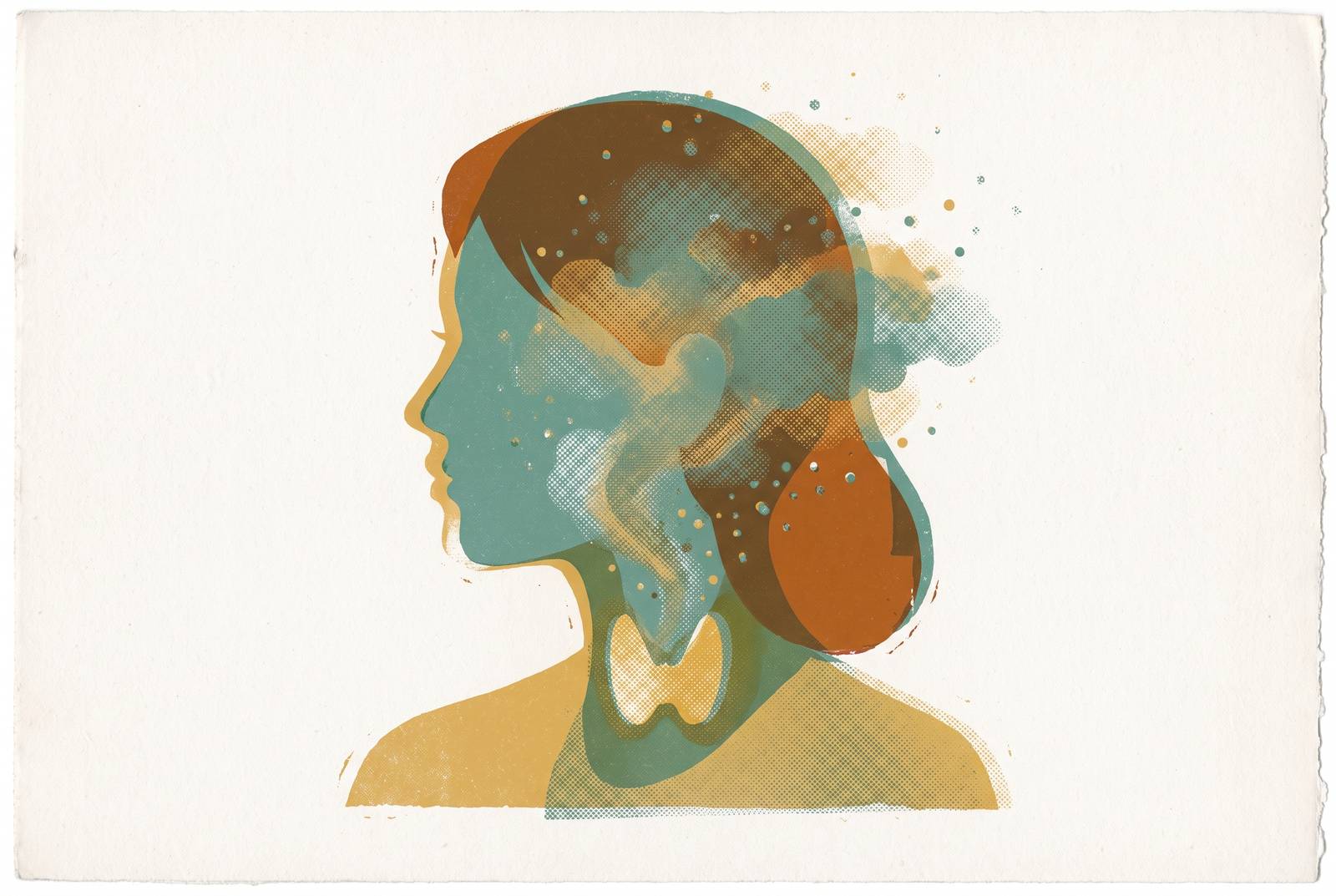
Thyroid dysfunction is a documented, physiologically direct cause of cognitive symptoms in women, and it belongs in the same diagnostic conversation as perimenopause, stress, and aging. Brain fog, memory lapses, and trouble concentrating are not automatically explained by "getting older" or "being busy." Sometimes they trace to a gland in the neck that most people only think about when something goes visibly wrong.
The story usually starts the same way. A woman in her 40s starts forgetting words mid-sentence, or walks into a room and cannot remember why. She mentions it to a clinician, who attributes it to stress, or anxiety, or normal aging, and moves on. Rarely does that conversation include a thyroid panel, even though the connection between thyroid function and cognition is well established in endocrinology and neurology literature. Part of the reason is structural: cognitive symptoms are subjective, they overlap across a dozen conditions, and no single specialty claims ownership of them. An endocrinologist thinks hormones. A psychiatrist thinks mood. A primary care physician, working with limited time, often defaults to reassurance. Meanwhile, roughly two-thirds of women report cognitive concerns during perimenopause in cross-sectional research, and globally, an estimated 50 million people live with dementia, with about 10 million new cases diagnosed every year. Thyroid dysfunction sits inside that landscape as a modifiable, testable risk factor, not a footnote. This piece traces the mechanism connecting thyroid hormone to brain function, distinguishes the cognitive signatures of an underactive versus an overactive thyroid, and walks through what a woman navigating unexplained cognitive change should actually ask for at her next appointment.
What thyroid hormones do in the brain
Thyroid hormones get filed under "metabolism" in most people's mental model, tied to weight and energy and not much else. That model is incomplete. T4 and T3, the two primary thyroid hormones, are directly active in brain tissue, not just circulating regulators of how fast the body burns calories.
T4 is technically a prohormone. It travels through the bloodstream in far greater quantity than T3, but it is not the active form the brain ultimately uses. Glial cells inside the brain convert T4 into T3 using an enzyme called type-2 deiodinase, so the brain does not simply receive active hormone passively from circulation. It manufactures what it needs, locally, from a precursor. That detail matters because it means brain-level thyroid hormone activity depends on more than what a blood test shows for T4 alone.
Once converted, T3 binds to thyroid hormone receptors, THRA and THRB, which are distributed widely across brain tissue, according to established endocrinology research indexed by the NIH's NCBI database. These receptors mediate T3's action at the cellular level, governing processes that sound abstract until translated into function. Thyroid hormones drive neurogenesis, the production of new neurons. They govern synaptogenesis, the formation of connections between neurons. They regulate myelination, the insulating sheath around neural pathways that determines how fast signals travel. They also direct how neurons and glial cells differentiate and migrate into position during development, and continue influencing plasticity afterward.
None of that is passive background chemistry. Thyroid hormones also shape the production and activity of serotonin, dopamine, and norepinephrine, neurotransmitters involved in attention, mood regulation, and memory consolidation. And there is a structural consequence to getting this wrong: research in endocrinology and neurology literature associates low thyroid hormone levels with reduced hippocampal volume, the hippocampus being a critical brain structure involved in forming new memories. So when a woman with an underactive thyroid says she cannot hold onto information the way she used to, that complaint has a plausible anatomical basis, not just a subjective one. The cognitive effects of thyroid dysfunction are not incidental. They follow directly from thyroid hormone's role in building and running the brain's circuitry.
How hypothyroidism impairs cognition: the slowed-signal profile
Overt hypothyroidism, meaning clearly low thyroid hormone confirmed by lab work, produces a fairly consistent cognitive signature. Memory loss, particularly in working memory (the mental scratchpad used to hold information temporarily) and verbal recall, appears first and most often. Attention becomes harder to sustain. Executive function, the set of skills involved in planning, sequencing tasks, and making decisions, weakens. Processing speed slows, motor coordination suffers, and lethargy sets in physically alongside the mental sluggishness. Neurology literature indexed on NCBI Bookshelf shows some patients also develop psychiatric features, including depression and, less commonly, bipolar-like mood symptoms.
The subjective term for much of this is brain fog, described clinically as trouble focusing, generalized forgetfulness, and a kind of mental haze that does not lift with rest. It is one of the most frequently reported complaints among people with hypothyroidism, and research on more than 5,000 people with hypothyroidism confirms brain fog as a real, measurable symptom cluster, not an exaggeration or a subjective impression with no biological grounding.
What complicates the picture further is subclinical hypothyroidism, a state where TSH runs elevated but T3 and T4 remain within the normal reference range. Patients in this category frequently do not receive a diagnosis or treatment at all, because the standard lab flag never triggers. Metabolic medicine literature shows subclinical hypothyroidism is still associated with impaired working memory, slower processing speed, and measurable memory impairment. This is clinically significant precisely because so many women are in this range and are told, in effect, that their thyroid is fine. Technically, it might be functioning within range. Functionally, the brain may still be underserved.
And here is the detail that tends to surprise people already being treated: normalizing TSH with levothyroxine (LT4) monotherapy does not guarantee that cognitive symptoms resolve. This remains an open clinical question in the endocrinology literature: patients on LT4 can still report cognitive symptoms even after their TSH reads normal, and it is unclear whether LT4 monotherapy alone fully restores cognitive outcomes over time. So a normal TSH on a lab report is not automatically the end of the story for a woman who still feels foggy. Because a normal TSH on a lab report is not automatically the end of the story for a woman who still feels foggy, it reframes what "treated" actually means in this context.
Hyperthyroidism's disruptive cognitive pattern
If hypothyroidism is a signal that runs too slow, hyperthyroidism is a signal that runs too fast, and both disrupt cognition, just through opposite mechanisms. A 2026 narrative review in Frontiers in Endocrinology, authored by Shi, Han, Zheng and colleagues, covers both clinical hyperthyroidism and its subclinical form, establishing that the thyroid-cognition relationship is bidirectional rather than a one-way street tied only to deficiency.
Where hypothyroidism produces sluggishness, hyperthyroidism tends to produce anxiety, restlessness, and racing thoughts, an excess of mental activity rather than a shortage. Attention does not slow down so much as it fragments, scattering across too many competing thoughts to hold focus on one. Difficulty concentrating also occurs here, but it stems from mental overdrive rather than mental slowdown, an important distinction for anyone trying to self-diagnose based on symptom lists alone. Hyperthyroidism also brings physical companions, tremor, heart palpitations, and disrupted sleep, all of which compound the cognitive impairment indirectly, since fragmented sleep alone is enough to degrade memory and attention in anyone, thyroid status aside.
Subclinical hyperthyroidism remains similarly contested territory. The Frontiers review notes that conclusions across existing research are inconsistent, varying by age group studied and by which neuropsychological tools researchers used to measure cognition. That inconsistency is a reason to take age into account when interpreting any single study, since thyroid hormone's cognitive effects appear to differ across life stages. It is a reason to take age into account when interpreting any single study, since thyroid hormone's cognitive effects appear to differ across life stages, a confounding factor relevant to any midlife woman trying to map a study's findings onto her own experience. The throughline across both directions of thyroid dysfunction: too little hormone and too much hormone both disrupt cognitive function, just by different routes, and both deserve investigation rather than dismissal.
Why thyroid disease is disproportionately a women's midlife issue
Thyroid disease is not evenly distributed across the population, and the imbalance is stark. A Frontiers in Endocrinology source estimates hypothyroidism affects between 4.8% and 25.8% of women in the general population, compared with 0.9% to 7.9% of men. The wide range reflects differences in how studies define the condition and which populations they sample, but even at the low end, the sex disparity is significant and consistent.
The most common driver behind this gap is Hashimoto's thyroiditis, an autoimmune condition in which the immune system infiltrates the thyroid gland with lymphocytes, gradually causing fibrosis and atrophy of the tissue. It is the dominant cause of hypothyroidism in countries with sufficient dietary iodine, and it occurs substantially more often in women than in men. Hashimoto's is widely recognized as common, with incidence peaking in midlife. First-degree relatives of someone with Hashimoto's face elevated risk, and twin studies suggest a meaningful heritable component.
Line up that age window against perimenopause, and the overlap is hard to miss. A woman developing autoimmune thyroid disease in her 40s is, statistically, quite likely also moving through perimenopausal hormonal shifts at the same time. A woman developing autoimmune thyroid disease in her 40s is, statistically, quite likely also moving through perimenopausal hormonal shifts at the same time, a common convergence of two overlapping hormonal timelines. Women with a family history of Hashimoto's, or with early symptoms, have a reasonable case for thyroid evaluation before waiting on a TSH threshold to be crossed.
Overlap between thyroid symptoms and perimenopause symptoms
SSM Health Women's Health data shows that by age 40, close to 65% of women report moderate or severe perimenopausal symptoms, and hypothyroidism affects up to 10% of women in that same decade of life. Two large, overlapping populations, moving through two conditions that look remarkably alike on the surface.
Brain fog and memory lapses appear across studies in anywhere from 26% to 95% of perimenopausal women, a wide range that itself reflects how inconsistently the symptom gets measured, and it is also a defining hypothyroid complaint. Fatigue occurs in both. Weight changes occur in both, though for different reasons: perimenopause shifts body composition, while hypothyroidism slows metabolic rate directly. Mood disturbance, sleep disruption, and difficulty concentrating (reported by 52% to 90% of perimenopausal women, depending on the study) round out a symptom list that is functionally identical for an ovarian or thyroidal underlying cause.
Why exactly does this happen so often in clinical practice? Picture a woman in her mid-40s describing fatigue, brain fog, and mood changes to her doctor. The path of least resistance is to attribute all of it to perimenopause and move on, because the age and the symptom list fit that story cleanly. But that same path can run in the other direction. A thyroid diagnosis might get made and treated in isolation, with nobody checking FSH or estradiol, leaving the perimenopausal component of her symptoms untouched. Either error leaves a woman only partially treated. And the two conditions are not mutually exclusive. A woman can have untreated thyroid dysfunction stacked on top of perimenopausal hormonal flux, producing cognitive impairment that is additive, since the two conditions are not mutually exclusive.
There is a neurological parallel worth noting here, one that reinforces just how physically real both experiences are. Research has documented neurological changes in both groups: perimenopausal women show altered brain activation during memory tasks, while hypothyroid patients show decreased cerebral glucose metabolism. Different hormonal source, similar neurological signature, and both are measurable with appropriate clinical testing. The overlap in symptoms is the exact reason testing for both belongs in the same appointment. It is the exact reason testing for both belongs in the same appointment.
What testing looks like and why a single TSH result tells an incomplete story
Standard thyroid screening in most clinical settings starts and stops with a TSH (thyroid-stimulating hormone) blood test. It is widely used because it is cheap and generally reliable as a first pass, but relying on it alone leaves real gaps.
Subclinical hypothyroidism, discussed earlier, can produce genuine cognitive symptoms while TSH is comfortably inside the normal range. A single TSH draw is also just a snapshot: it says nothing about fluctuation over the weeks or months before or after the blood draw. And a normal TSH does not rule out autoimmune thyroid disease at all, because thyroid antibodies, specifically TPO antibodies and thyroglobulin antibodies, can run elevated for years before TSH itself shifts out of range.
A more complete workup includes TSH, free T4, and free T3, the last of which matters specifically because of the conversion step from T4 into active T3 that happens in brain tissue, discussed in the first section. Thyroid antibody testing (TPO Ab and thyroglobulin Ab) belongs in that panel too, especially for women with a family history of Hashimoto's. Beyond the thyroid itself, FSH, estradiol, and progesterone deserve a place in the same round of bloodwork for a woman in her 40s presenting with cognitive symptoms, because thyroid testing and ovarian hormone testing answer different halves of the same question. Neither one alone tells the full story.
Tracking these values over time carries real value too, arguably more than any single result. Hashimoto's is progressive, so a normal thyroid panel at 42 offers no guarantee of a normal panel at 45. Perimenopausal hormone levels are dynamic in their own right, with estradiol fluctuating unpredictably during the perimenopausal transition, so a single measurement only captures one frame of a moving picture. Longitudinal testing reveals a trend line that a one-time snapshot simply cannot, distinguishing a stable baseline from a problem that is actively developing. Research on more than 5,000 people with hypothyroidism found that patients on LT4 can still report cognitive symptoms even after their TSH reads normal, which is precisely why ongoing monitoring, not just the initial diagnosis, deserves attention for women already on levothyroxine. None of these numbers mean much without someone qualified to interpret them in context. A lab report full of values is not the same thing as an answer, and turning it into one requires clinical judgment, not just a reference range printed next to a result.
Bringing thyroid dysfunction into the conversation with a clinician
A real gap exists between women experiencing these symptoms and women getting tested for them, and it runs in both directions. Many women leave appointments without a thyroid panel simply because nobody suggested it, and many clinicians do not initiate one unprompted when the presenting complaint is cognitive rather than physical. Closing that gap often comes down to what gets said out loud in the exam room.
Before an appointment, it helps to document specifics rather than generalities. Instead of describing "brain fog," note which tasks are affected, how often the symptom shows up, and how disruptive it actually is day to day. Track accompanying physical symptoms too, things like fatigue, weight change, cold sensitivity, shifts in heart rate, or hair loss, since these often point toward thyroid involvement specifically. Menstrual cycle changes matter as well, since documenting irregularity gives a clinician the information needed to stage perimenopausal status. Family history of thyroid disease, Hashimoto's in particular, should be stated explicitly rather than assumed to already be on file.
At the appointment itself, specificity in the request matters. Asking for "a full thyroid panel including TSH, free T4, free T3, and thyroid antibodies" gets a different response than asking to have the thyroid checked in general terms, which often results in an order for a single marker rather than the full panel. Framing FSH and estradiol as part of the same hormonal workup, rather than a separate, unrelated request, keeps the conversation focused on the full picture rather than two disconnected asks.
If a clinician dismisses the request outright, the research backing it is not thin or speculative. The 2026 Frontiers in Endocrinology review from Shi, Han, Zheng and colleagues, along with early symptoms, and the NCBI Bookshelf literature on hypothyroidism's cognitive effects, all establish this as an area with genuine clinical grounding, not a fringe theory. A woman bringing that context into the room is not being difficult. She is asking for the workup the evidence already supports.