Cortisol Rhythm Disruption in Perimenopausal Women
Hormonal shifts, not just stress, destabilize cortisol rhythms during perimenopause.
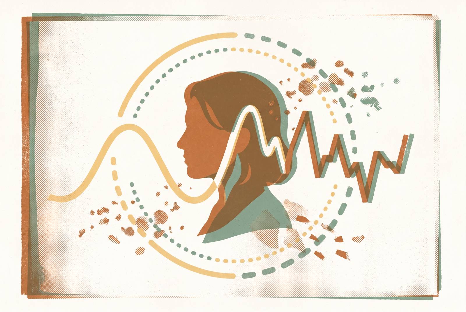
Cortisol rhythm disruption in perimenopausal women is not adequately explained as a stress or lifestyle problem. Changes in estrogen and progesterone can destabilize the hormonal system that governs cortisol, linking sleep, energy, mood, and metabolism. Understanding that mechanism explains why symptoms can cluster together, and why their timing can reveal more than a single hormone result.
How the healthy cortisol curve is supposed to work — and what estrogen has to do with it
Cortisol is a circadian hormone. Its daily rise and fall help organize metabolism, immune function, energy, and sleep. The suprachiasmatic nucleus, or SCN, in the hypothalamus acts as the master pacemaker, synchronizing peripheral clocks in nearly every cell, including the adrenal gland itself. Cortisol secretion therefore belongs to a coordinated timing system. Its healthy function depends on both when it rises and how much is present.
The normal curve peaks within the first hour of waking, a rise called the cortisol awakening response, or CAR. Cortisol then declines through the afternoon and reaches its lowest point in the late evening. That decline supports the transition toward melatonin’s nighttime rise and sleep. The two hormones work in reciprocal rhythms. The body needs both a strong morning activation signal and a quiet evening signal, each for a different purpose.
Estrogen regulates circadian gene expression and SCN sensitivity, helping coordinate clock signals with reproductive function. Changes in estrogen can therefore change circadian timing as well as metabolism, body temperature, and heart rate. The reproductive and stress-response systems are not independent. The hypothalamic-pituitary-adrenal axis, or HPA axis, which governs cortisol secretion, operates partly under reproductive hormonal regulation.
Why estrogen decline doesn't produce a clean, linear drop in cortisol regulation
The idea that estrogen simply “gets low” during perimenopause misses the transition’s defining instability. Under the STRAW+10 staging system, early perimenopause, Stage −2, can last several years while progesterone declines and estrogen remains erratic. Progesterone often falls first and stays low. Estrogen, meanwhile, may spike and then crash, so the regulatory system gets signals that swing up and down.
This distinction explains why symptoms can precede a low estrogen result. A fall from a high peak can be substantial even when the resulting measurement does not appear low. Vasomotor symptoms and sleep disruption can start during those swings. One estradiol measurement cannot describe that movement. Late perimenopause, Stage −1, often lasts one to three years and brings extended stretches without a period, more consistently low estrogen, and potentially rising FSH, but cortisol regulation may already have become disturbed.
Falling progesterone reduces allopregnanolone, weakening the receptor-mediated inhibition that normally restrains the HPA axis. With that brake reduced, the system can produce more cortisol with less provocation. External stress need not have increased at all. The distinction is between a greater burden on an unchanged system and a hormonal change that makes the system itself more reactive.
How the HPA axis becomes dysregulated when estrogen and progesterone lose their regulatory grip
The HPA axis begins with the hypothalamus releasing corticotropin-releasing hormone, or CRH. CRH prompts the pituitary to release adrenocorticotropic hormone, or ACTH, which stimulates the adrenal glands to produce cortisol. Cortisol then feeds back to the hypothalamus and pituitary, suppressing further activation. This is a negative feedback loop. Falling progesterone also reduces allopregnanolone-mediated GABA-A inhibition of CRH neurons, weakening another restraint on the cascade.
Cohn et al. (2023) used a GnRH agonist to suppress estradiol, experimentally mimicking the hormonal state of menopause, while examining the effects of sleep fragmentation. Sleep fragmentation raised bedtime cortisol and suppressed the morning CAR. Estradiol suppression produced a different result. It independently lowered bedtime cortisol and had no significant effect on CAR.
Hormonal change and sleep disruption can alter cortisol through distinct pathways, without moving every measurement in the same direction. An HPA axis that reacts more readily may produce higher baseline cortisol or stronger responses, while disturbed sleep can shift the daily curve toward more nighttime activation and less morning activation. Timing is the essential distinction. Elevated evening cortisol makes sleep onset difficult, whereas a blunted awakening response weakens the signal associated with morning alertness.
That leaves an important question open. How much of a particular woman’s altered curve reflects reproductive hormonal change, and how much reflects the sleep disruption developing around it?
The self-reinforcing loop — how disrupted sleep drives further cortisol dysregulation
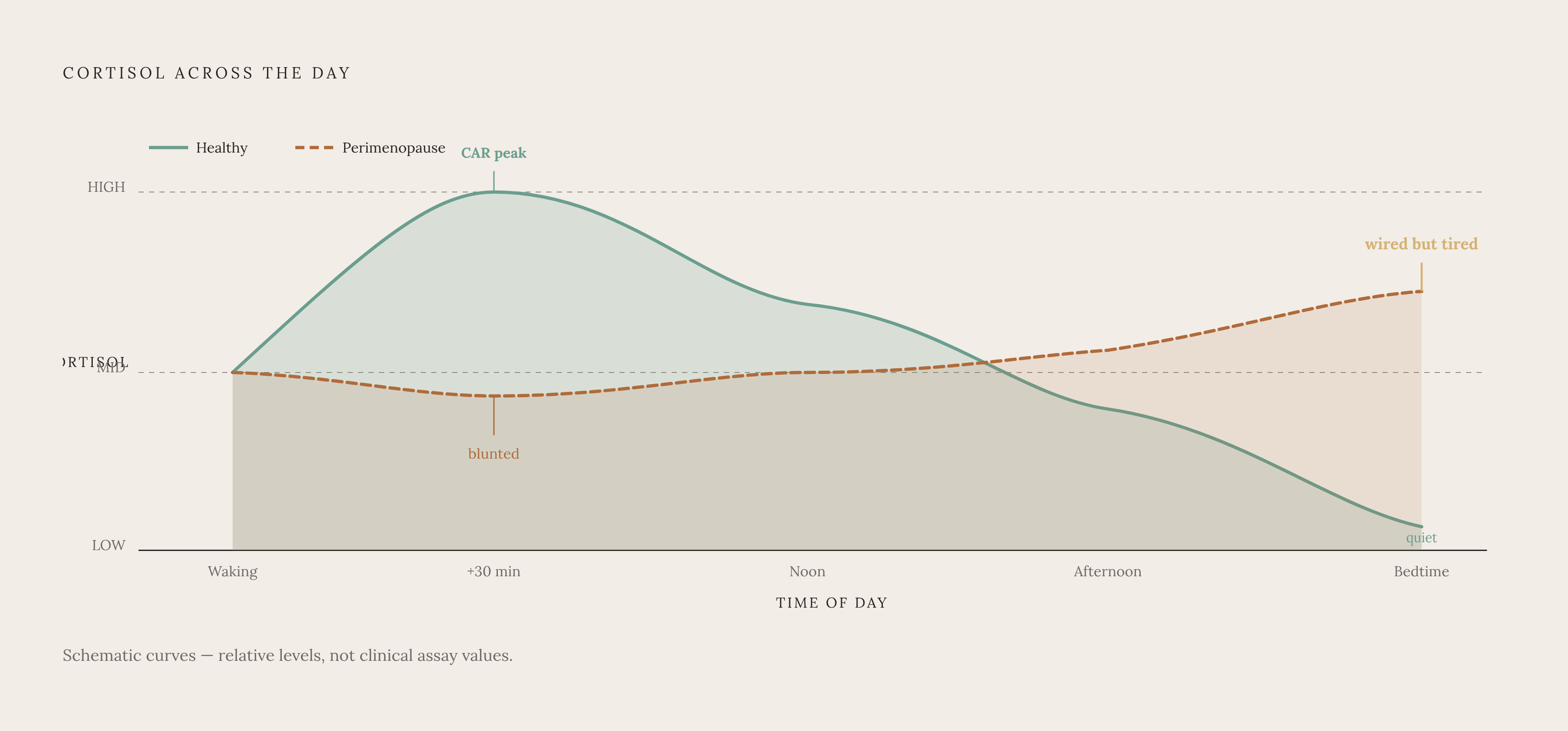
Once sleep is disrupted, it can become a driver of the cortisol disturbance that helped disrupt it. Deep sleep normally suppresses cortisol production. When sleep fragments and deep sleep is lost, that suppression grows weaker, so cortisol can stay high overnight. That nighttime activation then makes it harder to sleep again. The original hormonal trigger can thus set in motion a loop that no longer depends on that trigger alone.
The “wired but tired” pattern makes this timing conflict recognizable. Elevated evening cortisol supports arousal when sleep should begin, while a blunted morning CAR can leave the start of the day without its usual activation signal. Exhaustion and nighttime alertness reflect different parts of the same disrupted curve. Sleep disturbance is therefore both a consequence of dysregulation and a process capable of sustaining it.
Melatonin adds another vulnerability without replacing this explanation. Its levels decline with aging and the menopausal transition, reducing a sleep regulator independently of cortisol. When that decline coincides with elevated evening cortisol, the reciprocal signals that should favor sleep become less supportive. Both sides of the transition are affected. Less restorative sleep then feeds back into cortisol regulation, giving the disturbance a route to persist or intensify rather than settle after an isolated hormonal fluctuation.
Why cortisol rhythm disruption produces the symptom cluster perimenopausal women actually report
Why might abdominal weight redistribution happen along with anxiety, cognitive fog, fatigue, and night waking? Cortisol helps regulate several of the systems involved, so a disrupted rhythm can affect them together. Chronically elevated cortisol stimulates insulin release and repeated glucose surges. Over time, these changes can favor visceral fat accumulation and reduce insulin sensitivity. These metabolic effects are linked to cortisol dysregulation that lasts over time, while the healthy morning rise is needed for the body to function each day.
Loss of allopregnanolone’s receptor-mediated calming effect leaves the stress-response system with less inhibition, providing a biological route to anxiety and irritability. Life circumstances are not the only possible explanation. The elevated risk of a major depressive episode during perimenopause also warrants attention, although that risk does not establish cortisol disruption as the cause of every depressive symptom. Hormonal, sleep, and psychiatric processes can overlap.
Cognitive difficulty has two connected routes within this account. Chronically elevated cortisol affects the hippocampus, which supports memory consolidation and retrieval, while disrupted sleep independently impairs working memory, attention, and processing speed. Poor sleep adds its own cognitive burden. Brain fog, word-finding difficulty, and fatigue can therefore occur together as linked problems.
Vasomotor symptoms can further tighten these connections. Cortisol dysregulation can amplify hot flashes and night sweats, which can fragment sleep and raise cortisol further. This is why the complaints can cluster. They share regulatory pathways and can reinforce one another, although the presence of the cluster alone cannot establish which pathway is dominant in an individual woman.
Why a single cortisol test misses the problem — and what measurement actually captures the rhythm
A single cortisol measurement samples a point. A high value within the first hour of waking has a different meaning from a high value near midnight. Collection time is therefore essential to interpretation. Even a result taken at the right time cannot show whether the morning rise is blunted, the daytime decline is shallow, or bedtime cortisol stays high. Those questions require measurements across the day.
The Swiss Perimenopause Study followed 127 women aged 40–56 for 13 months and sampled salivary cortisol at months 2, 7, and 12. Its sampling times included waking, +30 min, +60 min, noon, and before bed, with an additional waking sample in month 2. This design captured both the awakening response and the diurnal slope. Testing again over time also showed how cortisol changed through the transition. It shows the kind of evidence over time needed to study rhythm, while leaving room for patients to follow different testing schedules.
Hair cortisol shows exposure over a longer time, while the daily pattern requires samples taken at different times within the day. FSH can fluctuate substantially and return to premenopausal levels after an elevated reading, while estradiol can change unpredictably. A normal snapshot does not necessarily explain fluctuating symptoms.
For women with irregular cycles, tests taken weeks or months apart may help show patterns when there is a clinical reason to repeat them. However, taking cortisol samples within a day and tracking reproductive hormones over a longer time answer different questions. One examines the daily rhythm. The other shows how hormones change through the transition, and a clinician needs to interpret either pattern to link it to symptoms.
Why cortisol rhythm disruption is so frequently missed, misattributed, or dismissed in clinical practice
Clinical recognition is difficult because several conditions can produce the same midlife complaints. Perimenopause and thyroid dysfunction overlap in fatigue, weight change, brain fog, mood shifts, cycle irregularity, and temperature intolerance. They can also occur together. Depression, anxiety, medication effects, and chronic stress add further possibilities, so a symptom pattern alone cannot reliably separate the causes. Menopausal changes can mask thyroid dysfunction, and thyroid dysfunction can obscure the menopausal transition.
This complexity makes a default explanation of stress tempting but insufficient. Woods et al. (2009) found that overnight cortisol elevation in perimenopausal women tracked with reproductive hormonal status. Perceived stress and social factors had little link to overnight cortisol once the hormonal indicators were taken into account. That finding challenges a purely behavioral explanation. It demonstrates why hormonal regulation must be included in the assessment.
When appointments are separate or assessments focus on symptoms, sleep, mood, and metabolic complaints may be considered apart from the hormonal transition happening at the same time. Conversely, attributing everything to perimenopause risks missing coexisting disease. Understanding cortisol timing gives clinicians a framework for asking which hormonal, sleep, and other clinical evidence would distinguish the competing explanations.
What understanding the mechanism means practically for a woman navigating perimenopause
For a woman navigating these symptoms, the practical shift is to bring their timing and relationships into the clinical conversation. Exhaustion, anxiety, weight redistribution, and night waking can be presented as a connected pattern. Could reproductive hormonal changes and disrupted cortisol timing be contributing, and what else needs to be considered? This framing leaves room for both a shared mechanism and coexisting conditions.
A woman can ask whether repeated, timed cortisol measurements would be useful and how a clinician would interpret them in relation to sleep and reproductive hormonal changes. She can also ask whether estradiol, progesterone, and FSH measurements would add useful context. More data are not automatically more informative. The value lies in selecting measurements that answer a clinical question and interpreting their changes.
Advice to stress less or sleep more cannot, on its own, explain a hormonal feedback system that has changed. Personalized, lab-based insight may help clarify that system when testing is appropriate by supporting a more specific conversation about what is driving the symptoms. Longitudinal insight makes self-advocacy more precise by allowing questions about when the disruption occurs, how it evolves, and whether the proposed explanation fits the pattern.
Sources
- Editorial: Circadian rhythm in adrenal endocrinology
- Cortisol Detection Methods and the Hormone’s Role in Evaluating Circadian Rhythm Disruption
- Mental health, sleep quality, and hormonal circadian rhythms in pregnant women with threatened preterm labor: a prospective observational study
- Sleep Disturbance and Perimenopause: A Narrative Review
- Perimenopause symptoms, severity, and healthcare seeking in women in the US