Muscle Loss and Anabolic Hormone Decline in Midlife Women
Hormone fluctuations in midlife trigger muscle loss before menopause officially begins.
A woman in her early 40s who trains the same way she has for a decade, eats roughly the same way, and still watches her strength slip and her waistline shift is not misreading her body. Hormonal changes begin well before the age most women associate with menopause, driving the symptoms women notice. Perimenopause can begin as early as the mid-30s, typically settles in during the 40s, and averages around four years, though it can stretch anywhere from two to a full decade. A study published in npj Women's Health, run jointly by UVA Health and the women's health app Flo Health, found that a majority of women between ages 30 and 35 already reported moderate to severe perimenopausal symptoms on the Menopause Rating Scale, a finding that upends assumptions about when symptoms begin. That is not a fringe finding.
The common mental model, menopause as a single event that arrives somewhere in a woman's early 50s, obscures the years of hormonal flux that precede it. Muscle loss occurs during the hormonal flux that precedes menopause, not as a downstream consequence after menopause finally lands. It is part of that earlier flux, running in parallel with the hormonal instability rather than trailing behind it. Understanding why requires looking at what is actually moving inside the body during those years, and how unevenly it moves.
Perimenopause and how hormone levels move through it
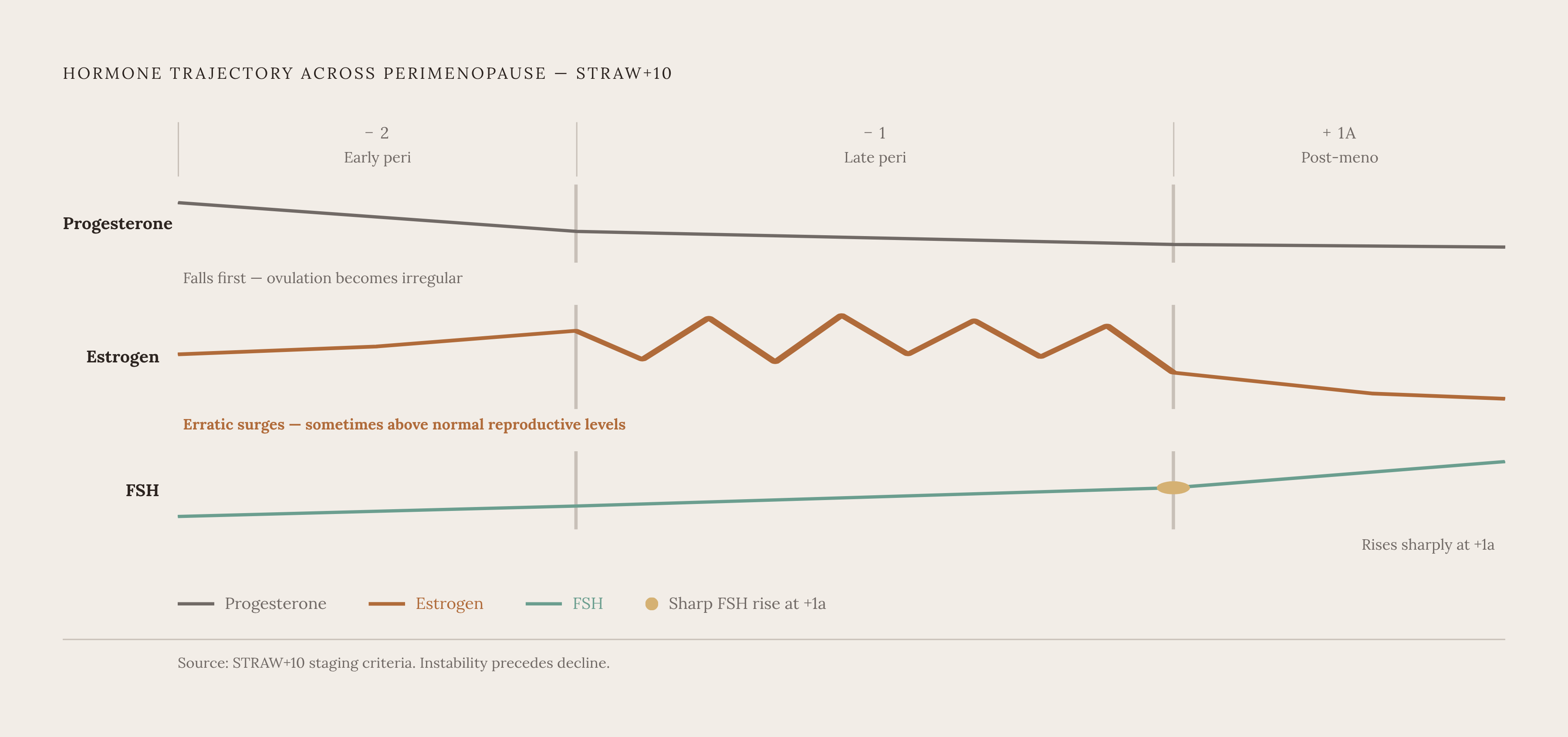
Perimenopause is a staged process in which hormone levels shift through distinct phases rather than sliding steadily from "normal" down to "low." It is staged, and each stage carries its own hormonal signature. The STRAW+10 framework, the standard clinical model for describing reproductive aging, breaks the transition into distinct phases rather than treating it as linear decline. In early perimenopause (Stage −2), progesterone starts declining first and estrogen turns erratic, with cycles becoming irregular by seven or more days. Only in Stage +1a, after a final period and a confirmed 12 consecutive months without bleeding, does FSH rise sharply and estrogen actually drop in a sustained way.
Notice what comes first: progesterone, not estrogen. Ovulation is what produces progesterone, and once ovulation becomes irregular, progesterone is the first hormone to fall off. Estrogen's story cuts against the popular shorthand of a woman "running out" of estrogen the way a car runs out of gas. Estrogen does not decline smoothly. It surges erratically through much of perimenopause, at times climbing higher than levels seen in a normal reproductive cycle, before it eventually declines in earnest. Most of what women experience as perimenopausal symptoms traces back to that instability, the whiplash of fluctuation.
This also contains an earlier signal that has not yet been identified. Inhibin-B, a hormone produced by ovarian follicles, starts declining and FSH starts rising before estradiol shows any dramatic shift. That means the earliest biological evidence of the transition often arrives before a woman or her clinician would think to look for it in estrogen alone.
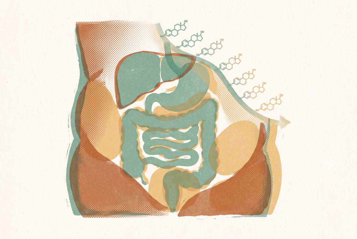
Androgens, testosterone included, continue a gradual decline that began after age 20, affecting energy, mood, and vitality. And cortisol tends to rise across this same period, because the reproductive hormone axis (HPG) and the stress hormone axis (HPA) are wired together; as estradiol and progesterone decline, the stress system responds in kind. None of these systems operate in isolation. That is precisely why the next question, what any of this does to muscle, cannot be answered by looking at estrogen alone. In late perimenopause, designated Stage −1, periods are absent for 60 or more days, vasomotor symptoms are likely, FSH is rising, and estrogen is still swinging.
How estrogen supports muscle tissue when it becomes unpredictable
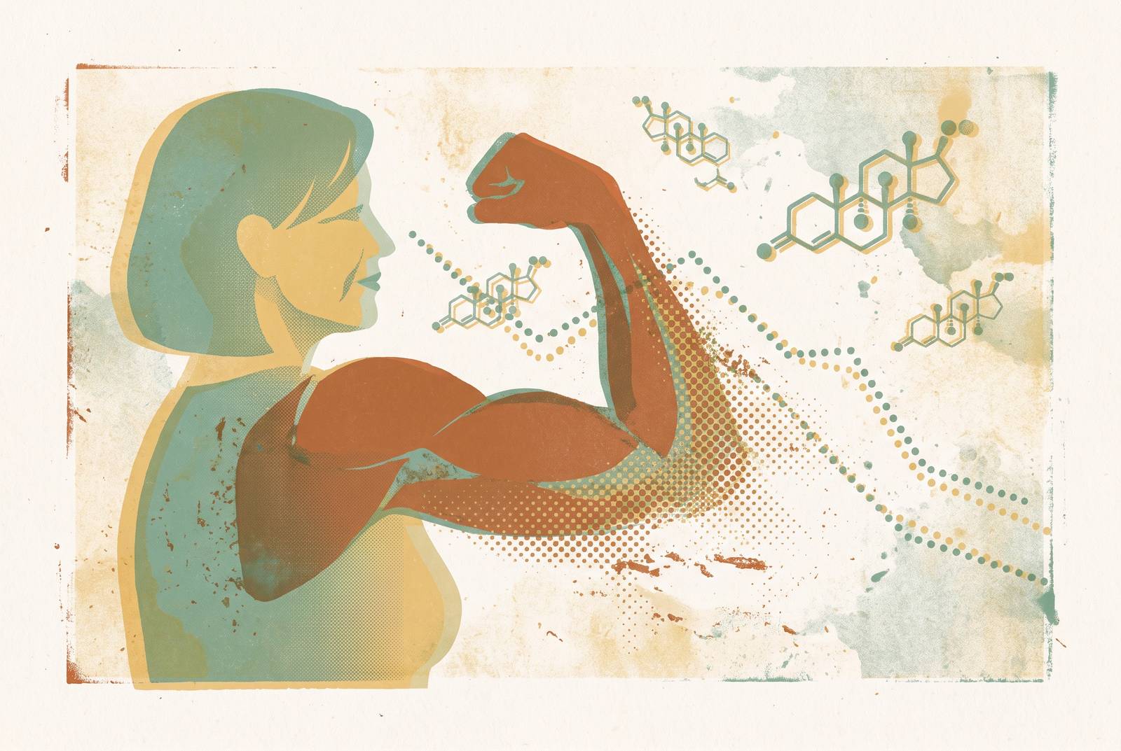
Estrogen is often filed under "reproductive hormone" and left there, but it behaves like an anabolic hormone too, one that plays a genuinely protective role in muscle tissue. When it drops, or simply becomes unstable, muscle becomes easier to lose and fat becomes easier to gain, particularly around the midsection. The quadriceps offer a concrete, almost literal illustration of this: they are among the first muscle groups to weaken during perimenopause, typically starting around age 45.
Why the quads specifically? They are unusually rich in fast-twitch muscle fibers, the fibers responsible for power output and quick reactive movement, the kind used in catching a stumble before it becomes a fall. Fast-twitch fibers appear especially sensitive to hormonal shifts. Without adequate estrogen support, they shrink faster than other fiber types and accumulate fat within the muscle tissue itself. That matters well beyond aesthetics. Weak quadriceps affect balance and bone loading, and strong quads are one of the better predictors of whether a woman maintains independent mobility after menopause or ends up at higher risk of falls and fractures.
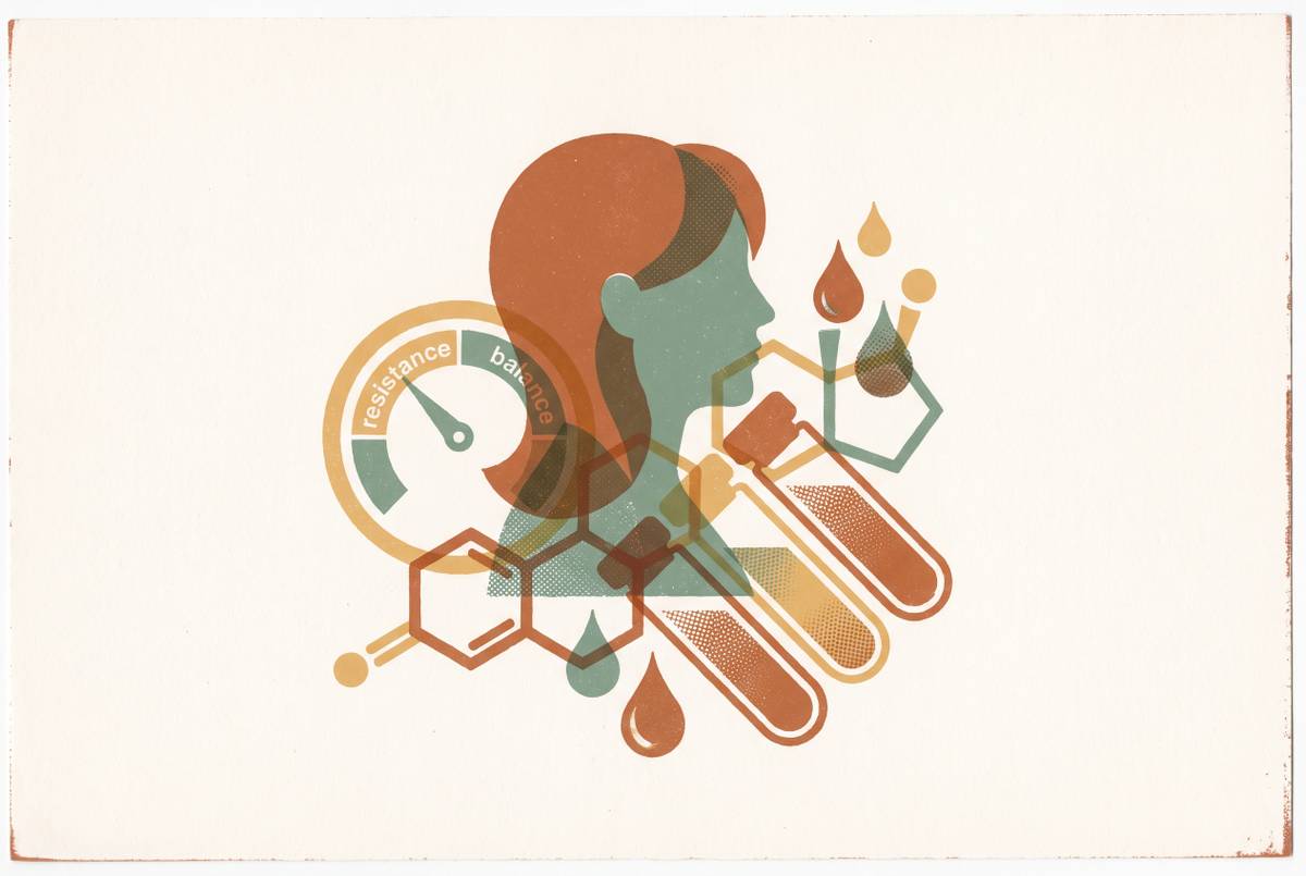
There is a second mechanism working against muscle at the same time, one that has nothing to do with fiber type: anabolic resistance. As estrogen declines, the body becomes less efficient at using dietary protein to trigger muscle repair and growth. More high-quality protein is required to produce the same anabolic signal that used to come from a smaller amount. Muscle protein synthesis, the biological process by which the body actually rebuilds muscle tissue in response to exercise or eating protein, appears blunted in older women in response to those stimuli, even when resting levels look entirely normal. In practice, that means the muscle-building machinery still exists. It has simply become less responsive to the exact triggers, resistance training and protein intake, that are supposed to switch it on.
A 2025 study in Frontiers in Endocrinology, led by Wang and colleagues at Shanghai Jiao Tong University, examined the relationship between menopause-related symptoms and muscle mass index directly. The study's own framing is notable: the exact mechanistic role estrogen plays in that relationship remains an active, open area of research. Which raises a fair question. If the mechanism is not fully settled, how confident can anyone be about the scale of the problem? That question deserves its own section.
What the research shows about how much muscle is lost (and what it cannot yet prove)
Here is where the story needs to slow down and get honest about its own limits, because an article that only asserts mechanism without checking it against the data is not worth much. A 2026 narrative review by Menzies, Bowtell, Shur, and Brook out of the University of Nottingham, published in the Journal of Cachexia, Sarcopenia and Muscle that February, pulled together the existing longitudinal and cross-sectional evidence on the question. The pattern held up reasonably well: four of five longitudinal studies and seven of eleven cross-sectional studies showed reductions in lean or muscle mass across the menopausal transition. The magnitude reported was a mean difference of about −2.5% in perimenopausal women and −5.7% in postmenopausal women, relative to premenopausal women.
A separate 2026 systematic review and meta-analysis by Kruse and colleagues, covering musculoskeletal manifestations of perimenopause across a very large sample of women and published in JBJS Open Access, stands as one of the primary sources worth reading in full on this question.
But how solid is the measurement that produces these numbers? Less solid than the percentages alone suggest. Most of this lean-mass evidence comes from DXA scans, dual-energy x-ray absorptiometry, which measures lean body mass broadly rather than skeletal muscle specifically, and may well underestimate the true change happening in muscle tissue. Techniques like MRI and methyl-D3-creatine dilution, a method that tracks a labeled creatine compound to estimate actual muscle mass, show stronger agreement with what is really going on in the tissue. That gap between what DXA sees and what is actually happening is not a minor footnote. It means the true deficit could be larger than the headline figures show, or it could be distributed differently than lean-mass numbers suggest.
There is also a harder gap: evidence on muscle protein breakdown across the menopausal transition is thin, almost absent, which makes it impossible right now to build a complete hormone-withdrawal story purely from human data. Wanting a clean, fully mechanistic account (estrogen drops, therefore muscle breaks down, full stop) runs ahead of what the science can currently support.
What the observational data does support is narrower, but still meaningful. A decline in that same free oestradiol index also tracked with declining handgrip strength. These are associations. Yet they point somewhere specific and consistent: hormones appear to relate more to muscle function and quality, strength and grip and power, than to raw bulk tissue mass. That distinction, function over volume, turns out to matter a great deal for what comes next.
Why a single hormone test gives an incomplete picture
If estrogen moves erratically rather than declining on a clean slope, then a single blood draw is trying to catch a moving target with one still photograph. The SWAN study, the Study of Women's Health Across the Nation, is one of the largest longitudinal projects tracking women through this transition, and it found that vasomotor symptoms, hot flashes and night sweats among the clearest outward signs of hormonal disruption, lasted a median of 7.4 years and fluctuated substantially from month to month within the very same woman. Symptoms swing that much over years. Why would anyone expect a single hormone number, drawn on a single day, to tell a stable story?
It doesn't, and the erratic surge-then-decline pattern of estrogen described earlier makes that concrete. A single estrogen reading could land high, normal, or low depending entirely on where in that erratic trajectory the blood happened to be drawn, and without knowing that context, the number by itself is close to uninterpretable. Layer onto that the earlier point about inhibin-B and FSH shifting before estradiol does. A woman or a clinician watching estradiol alone risks missing the earliest signals of the transition entirely, simply because they show up somewhere else first.
Clinical guidance needs to be read carefully here, because it is easy to misquote. The 2025 European Society of Endocrinology clinical guideline advises against routine biochemical testing to diagnose perimenopause or menopause in women over 45 who already present with compatible symptoms, on the reasoning that diagnosis in that group should rest on clinical history. That is a guideline about how to label a diagnosis in a clinical encounter. It is not an argument against tracking hormone levels over time to understand one's own trajectory, and treating it as the same thing would be a misreading. Repeated measurement over time is what actually reveals the pattern that a single reading structurally cannot: whether estrogen is swinging widely, whether FSH is trending upward, whether progesterone has already declined. One might argue a single test still has some value, as a data point. It does, but only as one point plotted against others; on its own, it is closer to noise than signal. The logical conclusion of everything the biology says about erratic hormone movement is that longitudinal data, not a single snapshot, is what turns a number into something a woman can actually act on, and even then, that data needs expert interpretation to mean anything clinically useful.
Practical and clinical steps women can take
None of this points toward a fix, exactly, but it does point toward levers, and toward a clearer sense of where effort actually pays off. Resistance training remains the most evidence-backed intervention for preserving muscle through this transition, more so than cardio alone, because cardio does not create the muscle protein synthesis response needed to push back against anabolic resistance. Protein intake requirements rise as resistance builds, and the practical target for most active women over 50 is around 25 to 30 grams of protein per meal, spread across the day rather than loaded into one sitting, since spacing appears to produce better results than concentrating it.
But mass on a DXA printout may be the wrong thing to chase in the first place. A 2026 editorial by Phillips suggests that menopause affects muscle quality and performance, strength, power, balance, resistance to fatigue, more than it affects raw tissue volume. Function is the more meaningful target. That reframing gets support from a mapping study by O'Bryan and colleagues (also cited in the Phillips editorial), which tracked neuromuscular function across the adult female lifespan and identified an accelerated decline, one centered in peripheral muscle rather than the nervous system, that lines up with the typical age of menopause onset. The goal, in other words, is maintaining neuromuscular function rather than defending a number on a printout.
There is a quieter benefit to all of this that has nothing to do with training programs. Knowing that quadriceps weakness, shifting body composition, and declining strength in the mid-40s may be hormonally mediated, rather than simply age or a woman "letting herself go," changes the conversation a woman can have with a clinician. Perimenopause remains chronically under-diagnosed and under-explained, and a woman who arrives with a symptom timeline and some longitudinal sense of her own hormonal pattern is in a stronger position to get a specific answer instead of generic reassurance. That, more than any single supplement or training split, is the practical payoff of understanding the mechanism at all: not a guarantee, but a sharper set of questions, and a better shot at getting real answers to them.
Sources
- Menopause, Female Sex Hormones, Skeletal Muscle Mass and Muscle Protein Turnover in Humans - PubMed
- Menopause and Muscle: Closer to Answers, but Significant Questions Remain - PMC
- www.frontiersin.org
- Association between menopause-related symptoms and muscle mass index among perimenopausal and postmenopausal women and the mediating role of estrogen levels - PMC
- Associations Between Female Sex Hormones and Skeletal Muscle Ageing: The Baltimore Longitudinal Study of Aging - PMC
- Menopause, Female Sex Hormones, Skeletal Muscle Mass and Muscle Protein Turnover in Humans - Menzies - 2026 - Journal of Cachexia, Sarcopenia and Muscle - Wiley Online Library