Neuroinflammation and Brain Fog in Perimenopause
Declining estrogen triggers brain inflammation and cognitive decline, not mood disorders.
Brain fog in perimenopause is not a mood problem or a symptom of aging that women are meant to shrug off. It is a documented, mechanistic response to declining and erratic estrogen, one that appears in blood work, is visible in brain scans, and occurs in the daily experience of forgetting a word mid-sentence or losing a thread of thought that was clear thirty seconds earlier. This piece traces that mechanism from the molecular level up through what shows up on an MRI, and asks what the science actually supports and where it still falls short.
The scale of the problem is not small. Estimates from El Khoudary and colleagues and Sullivan and colleagues, cited in a USU Extension review, put subjective cognitive decline during perimenopause somewhere between 44% and 62% of women. A Lancet personal view by Gurvich, Spector, and Hickey, summarized in Urban Health Today, puts the figure even higher in cross-sectional samples: roughly two-thirds of women report cognitive concerns during the transition, with memory complaints ranging anywhere from 26% to 95% depending on the study, and concentration difficulties reported by 52% to 90%. That range is wide enough to look messy, but a consistent pattern produces it, shown by the figures themselves holding steady in what they describe even as they vary from 26% to 95% and 52% to 90%. Brain fog isn't formally diagnosed the way, say, hypothyroidism is. The USU Extension piece describes it as a cluster: difficulty focusing, forgetfulness, slowed thinking. No lab test confirms it, no standard diagnostic code captures it cleanly, and that absence of a formal diagnosis creates a strange gap. Women describe something concrete and disruptive, and medicine, lacking a tidy category for it, often reaches for the nearest available explanation: stress, aging, mood. That reach is the actual problem here, and it is the first step toward taking the underlying biology seriously.
What estrogen does inside the brain, before it starts declining
Estrogen is not a reproductive hormone that happens to have some side effects on the brain. It crosses the blood-brain barrier freely and binds to receptors in regions including the hippocampus and prefrontal cortex, two areas most responsible for memory formation and executive function. Those aren't peripheral outposts. They're the control centers for exactly the kind of thinking that perimenopausal women report losing.
Estrogen also supports dopamine signaling, and dopamine is the neurotransmitter that underlies focus, motivation, and working memory. So when estrogen drops or swings unpredictably, dopamine-dependent processes wobble along with it, and attention problems occur or get worse. That's a fairly direct chain: hormone to neurotransmitter to cognitive function.
Less well known, and arguably more important for this discussion, is estrogen's role as an anti-inflammatory regulator inside the brain. It promotes what's called an anti-inflammatory glial phenotype, keeping the brain's immune cells, microglia and astrocytes, in a calm, reparative mode rather than an aggressive one. It downregulates pro-inflammatory cytokines like IL-1β, IL-6, and TNF-α. Microglia and astrocytes actually express estrogen receptors themselves, so this isn't an indirect effect. The hormone is talking directly to the immune cells living inside the skull.
Estrogen also supports neuronal energy demands and helps limit oxidative stress, which matters because neurons are energy-hungry cells with little tolerance for oxidative damage. It also plays broader protective roles in the neural environment, supporting the conditions neurons need to function and survive. Progesterone plays a parallel, complementary role: it has a calming, sedative effect through the GABA pathway, and its decline during perimenopause is a direct contributor to the sleep fragmentation and anxiety so many women report. The picture is a set of specific systems, immune regulation, energy metabolism, barrier integrity, neurotransmitter support, all losing a steady hand at once, not just "hormones go down."" It's a set of specific systems, immune regulation, energy metabolism, barrier integrity, neurotransmitter support, all losing a steady hand at once.
How Perimenopause Disrupts Estrogen Beyond Simple Decline
Here's where the popular understanding usually gets it wrong. Perimenopause is a period of wide, often erratic swings in estradiol, including transient spikes, before the overall trend turns downward. It's a period of wide, often erratic swings in estradiol, including transient spikes, before the overall trend turns downward. The instability is the hallmark, not the eventual low point.
The STRAW+10 staging framework, a widely used system for describing the menopause transition, breaks this into stages that make the volatility legible. In Stage -2, early perimenopause, progesterone is already declining and cycles are becoming irregular while estrogen bounces erratically. By Stage -1, late perimenopause, estrogen remains highly erratic and FSH (follicle-stimulating hormone) has already climbed substantially, sometimes reaching levels typically associated with full menopause, though still highly variable from one test to the next. It's only in Stage +1a, after the final menstrual period, that estradiol decline becomes more sustained, in the way most people picture when they think of menopause.
FSH itself deserves a closer look. It's traditionally treated as just a marker, a number that tells a clinician where someone sits in the transition. But emerging research, discussed in an S1 research brief referencing work on the APOE4-FSH axis, suggests FSH might be doing more than reporting on the process. It may actively activate signaling pathways implicated in amyloid and tau pathology, the same proteins associated with Alzheimer's disease. That reframes FSH from bystander to potential participant, though this is an emerging line of inquiry rather than settled fact.
Add cortisol to the mix. Cortisol, the main marker of HPA axis stress response, tends to rise during perimenopause too, and that rise adds further physiological stress to the instability already building from erratic estrogen. So a woman in late perimenopause isn't dealing with one destabilized system. She's dealing with several, moving at different speeds, occasionally in the same direction and occasionally not. The brain evolved to run on a fairly steady estrogenic signal. When that signal starts behaving erratically instead of just weakening, the whole system has to keep readjusting, which helps explain why symptoms can feel wildly different from one day to the next.
The neuroinflammation mechanism: what happens when estrogen's restraint on microglia is removed
Microglia are the brain's resident immune cells, and under normal circumstances, with estrogen doing its regulatory work, they sit in an anti-inflammatory, tissue-repairing mode. Think of estrogen as a hand on the brake. When that hand is steady, microglia clean up debris, prune unnecessary synapses, and generally keep the neural environment tidy.
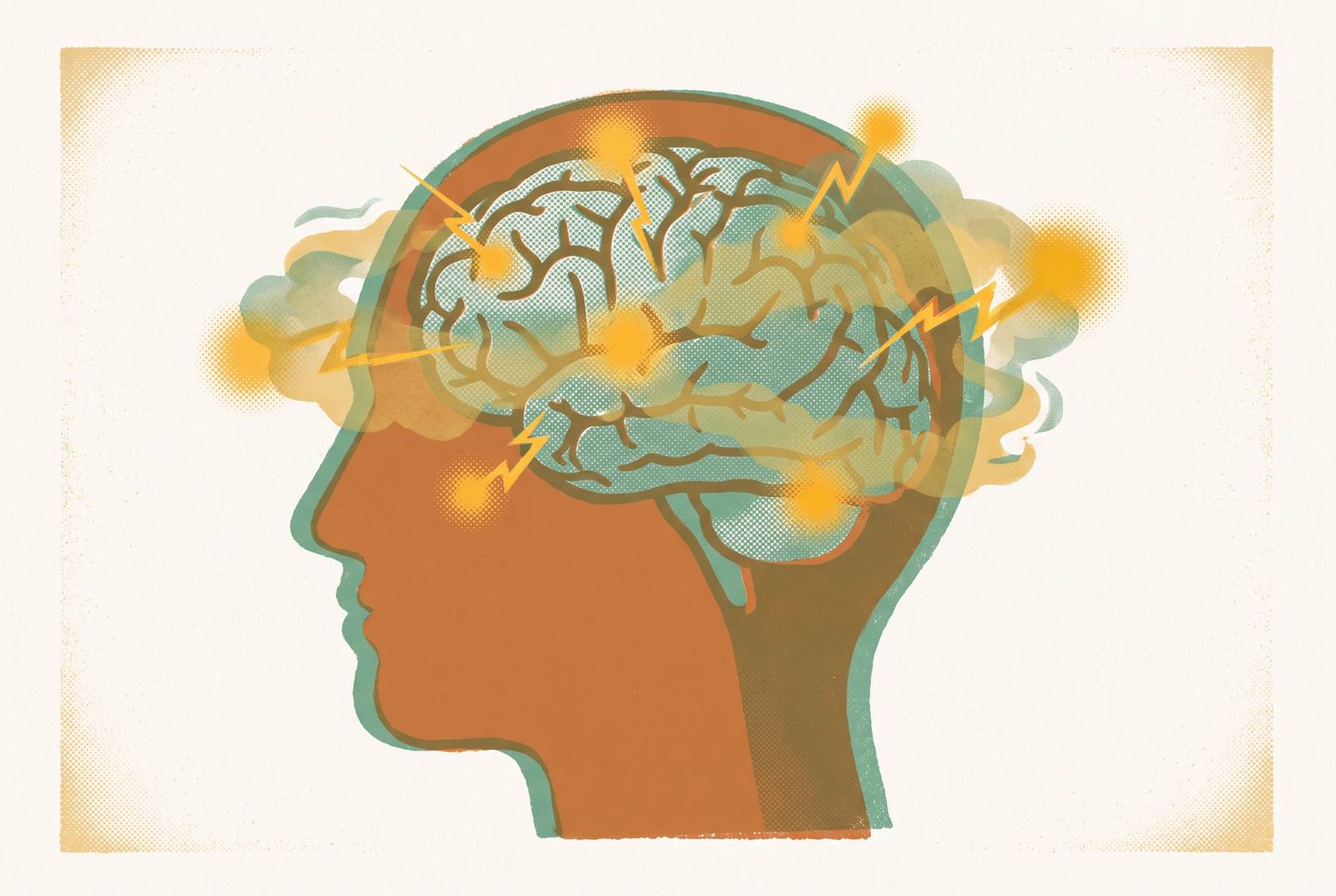
Remove the hand, or make it unpredictable, and microglia shift toward a pro-inflammatory state, moving away from their normal tissue-repairing role and contributing to chronic low-grade neuroinflammation. This accelerates chronic low-grade neuroinflammation, which is already recognized as a hallmark of brain aging even outside the context of menopause. So perimenopause, in effect, hits the accelerator on a process that would otherwise unfold much more slowly.
The downstream effects read like a checklist of what women actually report losing. Synaptotoxicity, meaning damage to the connections between neurons, disruption of blood-brain barrier integrity, mitochondrial dysfunction, and a kind of network disconnection where different brain regions stop communicating as efficiently. Each of these maps onto a specific cognitive complaint: word-finding trouble, slower processing, that sense of thoughts scattering before they can be grabbed.
APOE4, a gene variant associated with elevated Alzheimer's risk, impairs microglia's ability to clean up cellular debris (a process called phagocytosis), disrupts lipid handling and lysosomal function. APOE4, a gene variant associated with elevated Alzheimer's risk, impairs microglia's ability to clean up cellular debris (a process called phagocytosis), disrupts lipid handling and lysosomal function, and compromises the blood vessels that supply the brain. Estrogen normally works against these tendencies, offsetting some of the vulnerability APOE4 carriers face. Take estrogen's counteraction away during perimenopause, and APOE4 carriers may face a compounded risk, one layer of vulnerability stacked on another.
And the mechanism doesn't stop at memory and focus. The amygdala, hippocampus, and prefrontal cortex, the regions that govern anxiety and emotional regulation, sit among the areas most affected by neuroinflammatory signaling during hormonal transitions. Neuroinflammation is increasingly viewed as a central driver of the psychiatric symptoms, anxiety and depression, that spike sharply in late perimenopause. If the cognitive fog and the mood symptoms trace back to the same inflammatory pathway, clinicians so often treat them as separate complaints requiring separate explanations.
Longitudinal imaging from the Weill Cornell Mosconi Lab adds weight to this picture. Menopausal and perimenopausal women showed measurable declines on estrogen-dependent memory tests compared to men of similar age. The menopausal group in that research showed the highest rate of hippocampal volume loss, with statistical significance at p ≤.001, along with higher rates of amyloid-beta deposition compared to men. That's not a subjective report. That's tissue.
Brain Scan Evidence of Structural Changes Matching Women's Reports
The imaging data lines up with what women describe, and this pairing means these complaints aren't floating free of any physical correlate. Research presented at the 2025 Annual Meeting of The Menopause Society, held in Orlando from October 21 to 25, documented reductions in gray matter volume in the frontal and temporal cortices and the hippocampus, exactly the regions tied to memory and decision-making, and these reductions were linked to measurable declines in verbal and visuospatial memory performance.
White matter hyperintensities, which show up as bright spots on an MRI and indicate tissue damage tied to reduced cerebral blood flow, turn out to be more common during menopause, particularly for women who go through early menopause or who experience frequent hot flashes. Science Daily's coverage of the Menopause Society's October 2025 findings tied these hyperintensities to a higher risk of cognitive decline, balance problems, mood changes, stroke, and dementia. That's a fairly serious list of downstream associations for something that starts as a bright spot on a scan.
A University of Cambridge study published in Psychological Medicine found gray matter loss in regions tied to memory and emotional regulation, and many of the same women in that study also reported increased anxiety, depression, and fatigue. Structural change and subjective experience were tracking together, not diverging.
Population data drawn from the UK Biobank MRI subset, the Cam-CAN cohort, and the Tata Longitudinal Study of Aging suggests the pattern is regionally specific rather than a uniform, brain-wide shrinkage. Different cohorts implicate different structures, frontal gray matter in some, total gray matter volume in others, white matter hyperintensity fraction in still others. Timing matters too: women who go through spontaneous menopause at or before 49 show higher white matter hyperintensity fraction and lower cerebrovascular reactivity compared with women who go through menopause later.
The scale of some of this research is worth noting on its own terms. A JAMA Network Open study examined menopause age, cognitive decline, Alzheimer's onset, and brain volume trajectories using 2,603 women with cognitive data, 1,287 with neuropathology confirmed at autopsy, and 774 with serial 3T MRI scans. That's not a small pilot study. That's the kind of sample size that lets researchers actually draw longitudinal conclusions rather than guess from a snapshot.
But the picture isn't purely one of decline, and that nuance matters. Some evidence, per the Menopause Society review covered by Science Daily, suggests gray matter volume may partially recover after menopause, which could reflect the brain adapting and rewiring itself rather than simply losing ground permanently. There's also an odd, unresolved wrinkle: estrogen receptor density appears to rise during the transition, possibly as the brain's own attempt to compensate for falling hormone levels, but in some studies that elevated receptor density has actually been associated with worse memory outcomes, not better. Nobody has fully explained that tension yet.
Why objective tests often miss what women are experiencing
So if the scans show real structural change, why do standard cognitive tests so often come back looking fine? Longitudinal cohort data from studies like SWAN and the Avon Longitudinal Study show measurable declines in verbal learning and memory during the transition, yet objective neuropsychological test scores generally stay within normal ranges. That gap between what a woman reports and what a standardized test captures is one of the most consistent and least explained findings in this entire body of research.
A 2025 systematic review and meta-analysis covering 5,007 participants, summarized in the Lancet personal view carried by Urban Health Today, found a statistically significant link between subjective cognitive decline and objective learning efficiency specifically, but not for working memory or processing speed. The connection between subjective cognitive decline and objective learning efficiency is statistically significant, but not for working memory or processing speed, so it is not uniform across every cognitive domain a test might measure.
Gurvich, Spector, and Hickey, writing in The Lancet Obstetrics, Gynaecology, & Women's Health in 2026, proposed a clinical definition: self-reported impairment in one or more cognitive areas, without notable objective decline, that fluctuates and causes distress or functional impact, but without sustained interference in daily activities. That definition does real work. It draws a clear line between menopause-related brain fog and something like mild cognitive impairment or dementia. Brain fog, under this framing, is its own distinct thing, with its own trajectory, not a warning sign of something worse to come. It's its own distinct thing, with its own trajectory.
One might argue the field's biggest obstacle right now isn't the biology at all, but the vocabulary. Gazerani, writing in Frontiers in Human Neuroscience, points out that "brain fog" remains inconsistently defined and measured across studies, which limits how well findings compare to each other and constrains any real progress toward prevention-focused strategies. If every study measures a slightly different thing under the same name, comparing results becomes an exercise in translation rather than synthesis.
None of this means the subjective-objective gap is evidence that women's symptoms aren't real. It reflects, instead, the limits of standard cognitive tests, which were built to detect broad, stable impairment, not the kind of fluctuating, domain-specific, real-world difficulty that occurs when someone is trying to hold a complex thought together during a meeting while distracted by a hot flash. And the experience itself varies widely. Some women notice transient changes that pass. Others report minimal symptoms. A smaller subgroup deals with something persistent and functionally disruptive, a distinction laid out by Maki and Jaff in 2024 that's easy to lose in aggregate statistics.
The Value of Hormone Tracking Over a Single Test
Because estradiol swings so widely and nonlinearly through perimenopause, a single hormone measurement tells an incomplete story. One low reading might just catch a low point in a naturally erratic cycle. One normal reading might do the same in reverse. Neither one, alone, says much about what the brain has actually been experiencing over the preceding months.
A fuller hormone panel, covering estradiol, progesterone, FSH, LH, and DHEA-S among other markers, gives different pieces of the same puzzle: where a woman sits in the transition, and how the relevant systems are shifting relative to each other. FSH trending upward across several tests over time carries different clinical weight than a single elevated reading caught on one particular day, and given the emerging research on FSH as a potential direct actor in neurological signaling pathways, that kind of trend data has real interpretive value, not just diagnostic tidiness.
Cortisol tells a similar story. Watched in isolation, a single elevated cortisol reading doesn't say much. Tracked alongside declining estrogen across multiple points in time, a rising cortisol trend suggests a compounding neuroinflammatory load, a pattern that only becomes visible once there's more than one data point to compare.
That raises an obvious, practical problem: hormone testing isn't a routine part of most women's medical care, and the broader gaps in medical knowledge about female hormone conditions mean many women walk into a clinical appointment with no longitudinal data. They're asked to describe a fluctuating, months-long experience in a fifteen-minute visit, with nothing to hand the clinician except a description of how they feel that day.
Knowing where someone sits along the hormonal trajectory, rather than whether a single number falls "in range," lets a woman understand whether her cognitive symptoms line up with a period of peak estrogenic instability, and lets her track whether those patterns shift as she moves further into the transition. But the data alone isn't the whole answer. A stack of lab values means little without a clinician who can read the trend and put it in context. That interpretation is what turns a lab report into something a woman can actually act on.
What the Research Shows and Doesn't Show About Long-Term Cognitive Risk
This is the part of the story that calls for real precision, because it's easy to overstate. The Lancet Obstetrics, Gynaecology, & Women's Health article by Gurvich and colleagues, from 2026, is explicit: there's currently no evidence linking menopause-related cognitive symptoms to a heightened risk of Alzheimer's disease specifically, even though subjective cognitive decline as a broader category is associated with future cognitive decline risk in general populations. Those are two different claims, and conflating them does readers a disservice.
The biological mechanism described earlier, linking a genetic risk variant, estrogen changes, and immune cell activity in the brain, offers a plausible pathway for accelerated Alzheimer's pathology in carriers of that genetic risk variant during the estrogen deficiency of perimenopause. But a plausible mechanism, however well-supported by accumulating cellular and molecular evidence, is not the same thing as a settled clinical finding about any individual woman's risk. The framework has accumulating cellular and molecular support, but a plausible mechanism is not the same thing as a settled clinical finding about any individual woman's risk. The certainty some might want from it isn't there yet.
Hormone therapy research adds more texture, and some genuine complication, to this picture. A 2024 meta-analysis found no overall effect of menopausal hormone therapy on cognitive domain scores, though estrogen started close to the onset of menopause was linked to improved verbal memory specifically. Combined estrogen-progesterone therapy, by contrast, was associated with a modest decline in global cognition screening scores. A separate 2025 meta-analysis found no evidence that hormone therapy either raises or lowers dementia risk one way or the other. The "critical window" hypothesis, the idea that the timing of hormone intervention relative to menopause onset determines whether it helps, is a genuinely active area of research. It is not yet a settled clinical protocol that a doctor can simply apply.
What the research does support, with more confidence, is that brain fog is time-limited for most women. It typically lasts, at minimum, roughly a year, and for some women stretches to several years, generally resolving at or around menopause itself. And the brain shows real signs of neuroplastic adaptation, the partial gray matter recovery noted earlier isn't a fluke finding, it appears across multiple lines of research.
Gazerani's April 2026 piece in Frontiers in Human Neuroscience frames midlife, roughly ages 40 to 60, spanning the menopause transition, as a kind of measurement window: a period when brain health symptoms are most detectable and, potentially, most modifiable. That's a framing of opportunity rather than inevitability, and it's probably the most useful way to hold all of this evidence at once. The science is still moving. What's already established is enough to take the symptom seriously, and enough to argue for tracking it over time rather than waiting until something becomes a crisis.
Talking to a Clinician About Cognitive Symptoms
Because brain fog carries no formal diagnosis, precision in describing it becomes the tool that makes it legible to a clinician. Naming the specific domain affected, memory, concentration, processing speed, makes symptoms legible to a clinician in a way that saying "I feel foggy" does not." So does timing: when did it start, and does it fluctuate day to day, or has it been steadily getting worse without any relief.
That fluctuation question actually does real clinical work. Steadily worsening cognitive symptoms that don't ebb and flow call for a different kind of clinical workup than symptoms that rise and fall in step with sleep disruption or hot flashes, a distinction drawn out in the 2026 USU Extension review. If the fog tracks with a bad night's sleep or a rough stretch of vasomotor symptoms, that pattern itself is diagnostically useful information, and it's worth bringing to the appointment already organized rather than leaving the clinician to piece it together from a vague complaint.
Vasomotor symptoms, hot flashes and night sweats, deserve their own mention in that conversation, given how consistently they've shown up alongside white matter changes and cognitive complaints throughout the research already discussed. A woman who can walk into an appointment with a rough timeline, which symptoms cluster together, whether hormone levels have been tracked at all, and whether the pattern is stable or worsening, gives a clinician something to actually work with instead of a single data point pulled from one bad week. That's the throughline across all of this: brain fog in perimenopause has a mechanism, a set of visible structural correlates, and a research base that keeps growing. What it still lacks, mostly, is the kind of longitudinal, well-organized information that would let women and their clinicians meet each other with more than a guess.
Sources
- Brain Fog During Menopause: Understanding What
- Menopause-related brain fog as a midlife window in women's brain aging: toward ecologically valid measurement and digital phenotyping
- Brain Fog and Beyond: What Science Knows About Cognition During Menopause | Urban Health Today
- Brain fog during menopause? Here’s what’s really going on
- Frontiers | The estrogen–brain interface in neuroinflammation: a multidimensional mechanistic insight
- frontiersin.org
- menopause.org